
Your orthodontist keeps several diagnostic records, X-rays, photos, and films in the orthodontic records.
They are a valuable resource for orthodontists to:
- Examine the entire oral cavity (including the teeth and jaws)
- Develop an effective treatment plan
- Analyze facial growth patterns
At Loudoun Orthodontics, we take pride in keeping detailed orthodontic records for our patients, enabling us to keep a constant eye on your oral health.
Learn more about the different types of orthodontic records and how they can benefit your oral health!
Table of Contents
First Off, What are Orthodontic Records?
Prior to beginning your orthodontic treatment (for example, getting braces or aligners), your orthodontist collects an array of valuable documents. They help the orthodontist understand your teeth and jaw health and lend a valuable helping hand in planning your treatment.
The 8 Types of Dental Records
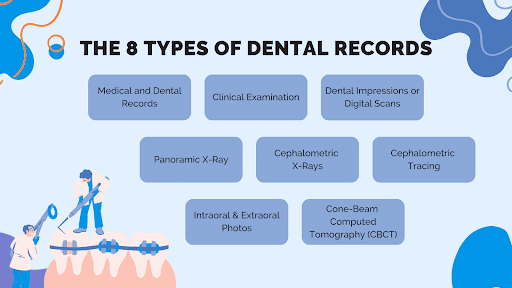
1. Medical and Dental Records
Patients’ medical and dental records contain information about their health, dental history, allergies, medications, as well as genetic conditions which may affect their teeth or facial appearance.
2. Clinical Examination
The purpose of a clinical examination is to thoroughly inspect the patient’s teeth, mouth, and jaw.
Before or during orthodontic treatment, the orthodontist assesses tooth alignment, spacing, and bite function, as well as any oral health issues.
3. Dental Impressions or Digital Scans
To create a 3D model of the teeth, the dentist takes an impression by having you bite into a soft material, which then hardens.
Today, creating a 3D digital model using a scanning process is more common.
Orthodontists can use either method to analyze the patient’s bite and devise a more effective treatment plan.
4. Panoramic X-Ray
Panoramic X-rays, also known as Panorex or simply “pano”, are a broad view of the whole oral cavity in one single image.
Dental and orthodontic professionals use this type of X-ray because it provides a 360-degree view of the oral cavity, allowing them to detect problems that might not be apparent otherwise.
A panoramic X-ray can detect impacted teeth, bone abnormalities, cysts, solid growths (tumors), infections, and fractures.

5. Cephalometric X-Rays
Cephalometric X-rays, commonly called “cephs,” are specialized radiographic examinations that capture a complete image of a side of the face.
In contrast to regular dental X-rays, cephalometric X-rays provide images of the entire side of the head, not just the teeth. They are invaluable tools for planning and evaluating orthodontic treatment because they reveal the relationship between the teeth, jaw, and facial profile.
Orthodontics uses cephalometric X-rays primarily to assess jaw growth and development relative to the skull. This information is particularly important when planning orthodontic treatment for young people. It is possible to determine whether the patient’s jaw is aligned properly or whether an overbite or underbite exists by taking an X-ray.
6. Cephalometric Tracing
Orthodontists use cephalometric tracing to determine a patient’s anatomical structure and plan treatments based on it.
When a cephalometric X-ray is taken, the film is covered with a clear sheet. It is then the orthodontist’s responsibility to trace the skull, jawline, teeth, and other relevant facial structures with a special pencil or digital tool. By tracing these points and lines, the patient’s craniofacial profile is created.
Cephalometric tracing is especially valuable in cases involving major structural changes to the jaw or face, such as orthognathic surgery, severe malocclusions, or orthodontic treatment planning for children or adolescents.
7. Intraoral & Extraoral Photos
Intraoral and extraoral dental photographs are used by orthodontists to assess, diagnose, and plan treatment strategies. They may also be used to document your treatment progress and to maintain comprehensive dental records.
An intraoral photo allows the orthodontist to capture detailed images of teeth and the surrounding structures, such as the gums, palate, and oral tissues. A mirror can help the orthodontist see hard-to-see areas like the back teeth, the inner surfaces of the upper and lower teeth, and the roof of the mouth.
A clear before-and-after intraoral photograph enables your orthodontist to observe the effectiveness of your orthodontic treatment. Such photos are particularly useful for identifying tooth decay, gum disease, crowded teeth, and other orthodontic issues.
Extraoral photos are taken from the outside of the mouth. They may include close-ups of the mouth, full-face photos, and profiles of the face.
By comparing the facial appearance with the underlying skeletal and dental structures, these pictures aim to show how these structures function. An extraoral photo can, for example, reveal whether the teeth are aligned with the facial structure, whether the jaw is imbalanced, or whether the smile is symmetrical.
8. Cone-Beam Computed Tomography (CBCT)
Dental and orthodontic imaging has been transformed by cone-beam computed tomography, or CBCT. This is an X-ray machine that produces 3D images of teeth, soft tissues, nerve pathways, and bone all at the same time.
CBCT provides a comprehensive, detailed image of the mouth, making precise diagnosis and treatment planning possible.
With CBCT, a series of images are taken from various angles around your head and then processed and rendered into a single 3D image by using special software. This comprehensive image can be manipulated on a computer screen, allowing the orthodontist to view oral structures from different angles and even zoom in on specific areas of interest.
The 3 Advantages of Cone-Beam Computed Tomography Systems Over Traditional X-Rays
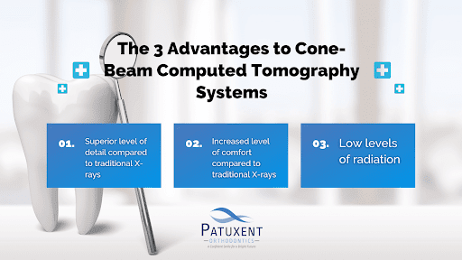
The first benefit of CBCT is that it provides a much higher degree of detail than traditional X-rays. It provides a complete three-dimensional view of the craniofacial structure, which is crucial for planning complex treatments such as orthognathic surgery, implant placement, and even managing impacted teeth.
In addition, CBCT scans are much faster and more comfortable than traditional dental X-rays. The scanning process is completed in less than a minute, and the patient does not need to bite down on any plastic holders.
The amount of radiation involved in CBCT is lower than that of a medical CT scan, which makes it a safer procedure for patients. However, it’s still primarily used in more complex cases, not for routine orthodontic exams.
How Do Orthodontic Records Benefit Your Treatment Plan?
Orthodontic records are the blueprint that guides the orthodontist in formulating the best course of action for achieving a patient’s dental and aesthetic goals.
Here are the 3 ways these records can benefit your orthodontic treatment:
Confirming a Diagnosis
Orthodontic records provide a detailed picture of your oral health status. This includes information about teeth position, tooth health, gum health, and the relationship between teeth and jaws.
This detailed picture is essential for diagnosing and planning your orthodontic treatment — no orthodontist would be able to effectively diagnose and plan without them.
Determining Facial Growth Patterns
Orthodontic records are crucial in forecasting how your dental structure might change in the future.
Growth prediction is an integral part of orthodontic treatment for younger patients. By analyzing records, orthodontists can predict how a child’s bite and teeth will develop over time and determine the appropriate course of action.

Measuring the Progress of Your Orthodontic Treatment
Last but not least, orthodontic records help treatment providers track your progress over time.
If your orthodontist compares current records with those taken earlier in the treatment process, they can determine whether the treatment plan is on track.
With this knowledge, any issues can be detected early on, and your treatment can proceed as planned!
How Long Must an Orthodontic Office Keep Patient Records?
An orthodontic office must keep patient records for a variety of reasons, including:
- Local and state laws
- The type of record
- The age of the patient
- The guidelines set by professional orthodontic associations
Orthodontic offices must be familiar with these regulations in order to remain in compliance and effectively manage their records.
Local and State Laws Vary Widely
Local and state laws require that dental records be kept for a minimum of 7 to 10 years from the date of the last treatment.
Records may be kept longer if the patient is a minor. For example, if the patient was under 18 at the time of treatment, records would typically be kept until the patient reached the age of majority (18 years of age in 48 states) plus the standard record retention period (7-10 years).
American Association of Orthodontists (AAO) Guidelines
AAO guidelines specify that patients’ complete patient records should be retained for at least 10 years from the date they were last seen, or until they turn 25 years of age.
Some Orthodontic Records are Kept Longer Than Others
Certain types of records must be preserved for a longer period of time than others. For example, radiographs and study models, which can provide important diagnostic and treatment information, may require indefinite preservation.

Start Your Orthodontic Treatment at Loudoun Orthodontics!
Contact Loudoun Orthodontics if orthodontic care is the solution to your dental woes. Whether you want to learn more about the benefits of orthodontic care or have questions about the process, use our live chat or call (703) 858-0303 or send us a message through our contact us page to connect with our friendly staff today and book a complimentary orthodontic consultation!
Our office, located at 19465 Deerfield Ave. Suite 304 Lansdowne, VA 20176, proudly serves the Lansdowne, VA area. So, if you’re residing in Ashburn, Sterling or Leesburg, and are looking for one of the best orthodontists in Virginia, don’t hesitate to visit our office!
We also invite you to keep up with our blog to get answers to many of the frequently asked questions about maintaining your perfect smile and follow us on Facebook and Instagram to become a part of our smiling community!
References
- Bakker, Mischa, and Joyce Miller. “Does an Audible Release Improve the Outcome of a Chiropractic Adjustment?” The Journal of the Canadian Chiropractic Association, Sept. 2004, www.ncbi.nlm.nih.gov/pmc/articles/PMC1769448/. Accessed 26 Jun. 2023.
- “Aao Home.” American Association of Orthodontists, aaoinfo.org/. Accessed 26 Jun. 2023.